Image
Width
2100
Height
1848
Permitted usage:
Public
Date added:
File size:
356.53 KB
MIME type:
image/jpg
About this file:
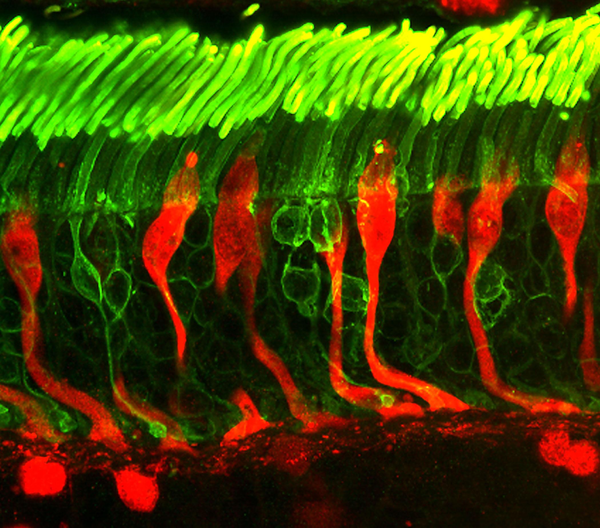
This is a confocal microscope image of rod and cone photoreceptors in a human retina. Fluorescent probes have been used to identify rod photoreceptors (green) and cone photoreceptors and horizontal cells (red). The cones looks like candle sticks, warped as if viewed through a fun house mirror. The horizontal cells are the round cells in the lower portion of the image.
Image courtesy of Dr. Robert Fariss, National Eye Institute, NIH
This image is licensed as U.S. Government Works, see https://www.usa.gov/government-works