Image
Width
994
Height
991
Permitted usage:
Public
Date added:
File size:
1.28 MB
MIME type:
image/png
About this file:
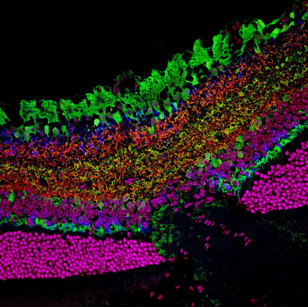
Confocal micrograph of optic nerve head region in adult mouse retina. The micrograph depicts staining for bipolar cells with an antibody against PKC- (blue), staining for horizontal cells and amacrine cells with an antibody against calbindin (green) and synapses of amacrine cells in the inner plexiform layer of the mouse retina with an antibody against synapsin (red). Nuclei are shown in magenta. Image was captured under 40x magnification.
This image is licensed as U.S. Government Works, see https://www.usa.gov/government-works