Image
Width
825
Height
823
Permitted usage:
Public
Date added:
File size:
1.82 MB
MIME type:
image/png
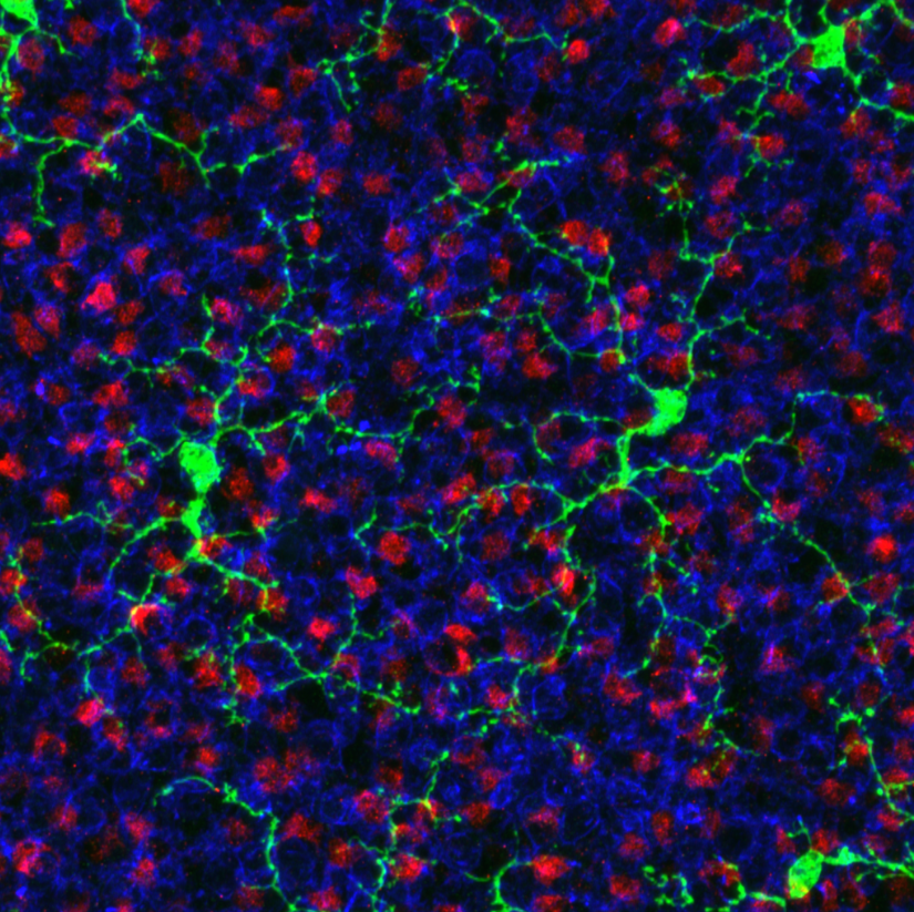
About this file:
En face view of GFP-expressing microglia (green) in the retina of a 2 month- old adult CX3CR1GFP/+ transgenic mouse. Presynaptic axon endings from cone photoreceptors (i.e. cone pedicles) labelled with cone arrestin (red) and postsynaptic rod bipolar dendrites labelled with PKCα (blue) are visualized in the same plane of the retina.
This image is licensed as U.S. Government Works, see https://www.usa.gov/government-works