Width
916
Height
938
Permitted usage:
Restricted
Date added:
File size:
148.55 KB
MIME type:
image/jpg
About this file:
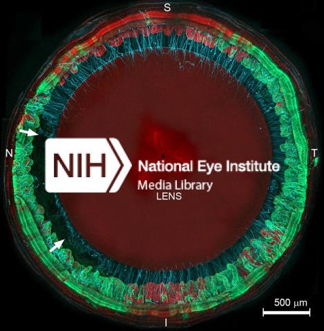
Cross-section of mouse eye showing lens, non-pigmented ciliary epithelium (green with sections of red at 12 o’clock and 6 o’clock), and zonular fibers (blue). Green corresponds to fibrillin-depleted NPCE. The space between the arrows indicates where the zonular fibers have snapped. Credit: Steven Bassnett.
This image is licensed as U.S. Government Works, see https://www.usa.gov/government-works
