Width
564
Height
564
Permitted usage:
Public
Date added:
File size:
52.89 KB
MIME type:
image/jpg
About this file:
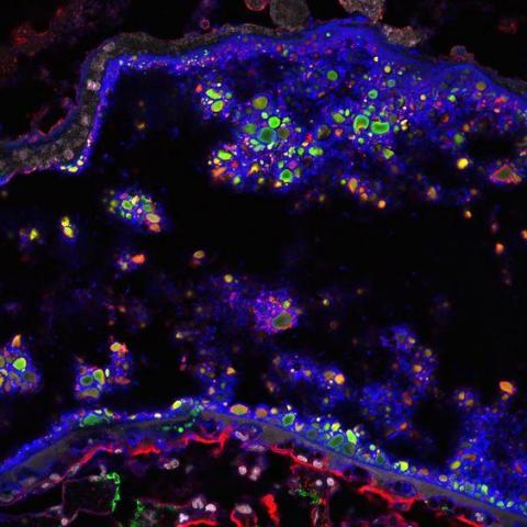
This is image of a human retina is from a person with AMD. Fluorescent probes have been used to identify different cellular and extracellular components in this section of tissue. Aggregates of fibulin-3 (green) and complement factor H (red) accumulated in lipid-rich deposits called drusen. Lipids are labeled with a fluorescent dye called filipin (blue). Individual cell nuclei appear gray in this image. Image courtesy of Robert Fariss, Mercedes Campos and Graeme Wistow, NEI.
This image is licensed as U.S. Government Works, see https://www.usa.gov/government-works
