Image
Width
564
Height
564
Permitted usage:
Public
Date added:
File size:
70.67 KB
MIME type:
image/jpg
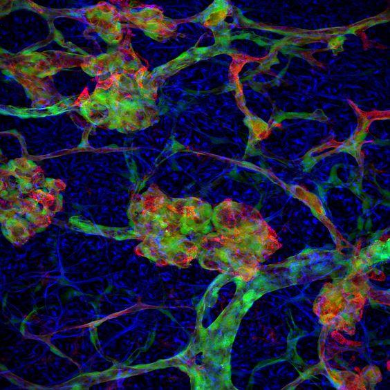
About this file:
This image shows blood vessels in the retina of a mouse. The vessels are abnormal due to exposure to a high concentration of oxygen shortly after birth. This model replicates a condition called retinopathy of prematurity. Vascular cells called pericytes (green) are closely associated with another class of cells called endothelial cells (red). Astrocytes, are labeled with an antibody to glial fibrillary acidic protein (blue). Courtesy of Robert Fariss and Jen-Yue Tsai, NEI.
This image is licensed as U.S. Government Works, see https://www.usa.gov/government-works